The anterior interventricular branch of the left coronary artery runs in the sulcus. The posterior interventricular sulcus.

Posterior Interventricular Artery An Overview Sciencedirect Topics
Is a groove between the ventricles on the back of the heart.

. Anterior and posterior venae cavae Internal. Which heart groove travels between the atria and the ventricles a anterior interventricular sulcus b. The other groove is the anterior interventricular sulcus situated on the sternocostal surface of the heart close to its left margin.
Vessel that follows the interventricular sulcus on the anterior surface of the heart and flows along the coronary sulcus into the coronary sinus on the posterior surface. Figure 5 illustrates anterior and posterior views of the surface of the heart. Called also anterior interventricular groove.
Posterior interventricular sulcus d. Once the vein reaches the left margin of the heart it circumvents to the posterior side and eventually converges with the oblique vein of the left. Is a valve in the interventricular septum that closes at birth.
The main arteries that run along the interventricular sulcus are the left anterior descending artery and the posterior interventricular artery. The other the anterior interventricular sulcus runs along the line between the right and left ventricles and contains a branch of the left coronary artery. Medical Definition of interventricular sulcus.
Asked Sep 27 2015 in Anatomy Physiology by Dylan. These grooves extend from the base of the ventricular portion to a notch the incisura apicis cordis on the. Other articles where anterior interventricular sulcus is discussed.
The anterior interventricular sulcus is situated on the sternocostal surface of the heart close to its left margin. The anterior interventricular sulcus is situated on the sternocostal surface of the heart close to its left margin. The anterior interventricular sulcus or anterior longitudinal sulcus is one of two grooves that separates the ventricles of the heart the other being the posterior interventricular sulcus.
This problem has been solved. The posterior interventricular artery supplies blood to the hearts posterior or bottom portion. Contains the great cardiac vein and coronary sinus.
Is another name for the coronary sulcus. Starting from the apex of the heart and running parallel with the anterior interventricular artery the great cardiac vein travels up along the anterior interventricular sulcus towards the base of the left atriums auricle. The crux of the heart is the meeting point of the interatrial and interventricular septa of the cardiac chambers.
Posterior interventricular sulcus one of the two grooves that separates the ventricles of the heart near the right. Coronary sulcus both a and b e. What is the function of the posterior interventricular artery.
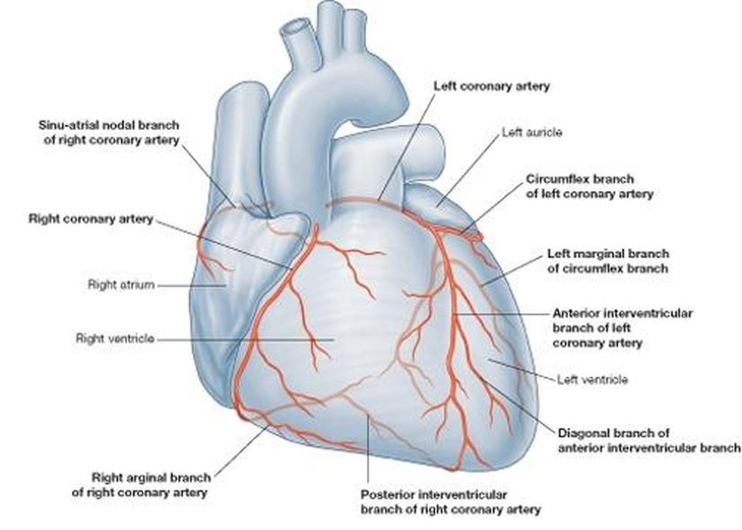
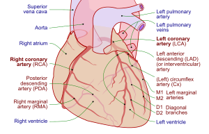
Coronary artery anatomy varies from person to person. Anterior interventricular sulcus one of two grooves that separates the ventricles of the heart near the left margin. Several large coronary branches emerge from this crown supplying blood to different parts of the heart.
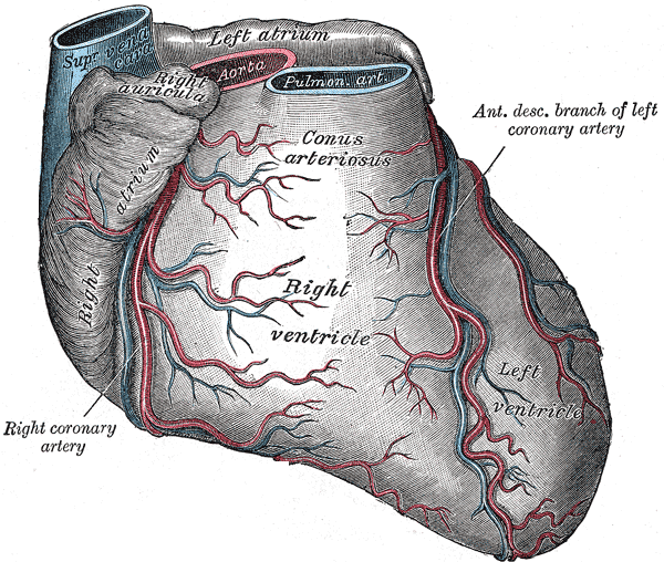
From Wikipedia the free encyclopedia. Inside the pericardium the surface features of the heart are visible. The ventricles are separated by two grooves one of which the anterior longitudinal sulcus is situated on the sternocostal surface of the heart close to its left margin the other posterior longitudinal sulcus on the diaphragmatic surface near the right margin.
Clinical Significance The coronary arteries can vary with respect to their origin number and course and many variants have been described in medical literature. The anterior interventricular sulcus or anterior longitudinal sulcus is one of two grooves that separates the ventricles of the heart the other being the posterior interventricular sulcus. Either of the anterior and posterior grooves on the surface of the heart that lie over the interventricular septum and join at the apex.
November 22 2021 Nora Advices. External surface of the heart. The epicardium and the 4.
Large vein that is in posterior aspect of coronary sulcus - collects blood and drains away from heart wall directly to the R. Sulcus interventricularis anterior TA anterior interventricular sulcus. Is a valve in the interventricular septum that closes at birth.
Asked Aug 29 2019 in Anatomy Physiology by Jezebella. The anterior interventricular sulcus is visible on the anterior surface of the heart whereas the posterior interventricular sulcus is visible on the posterior surface of the heart. The anterior interventricular sulcus is situated on the sternocostal surface of the heart close to its left margin.
Right and left atria 2. Interventricular groove may refer to. The posterior interventricular artery descends in the interventricular sulcus towards the apex of the heart.
The anterior interventricular vein is the largest and most consistent of the cardiac veins. A b and c 3. Parallels the anterior interventricular artery and drains the areas supplied by this vessel heart block interruption in the normal conduction pathway heart bulge.
When it reaches the apex it anastomoses with the anterior interventricular artery a branch of the left coronary artery. Called also interventricular groove. Right and left ventricles 3.
The phrenic nerve may lie close to the lateral. The _____ is in the anterior interventricular sulcus which drains the areas of the heart supplied by the left coronary artery left and right ventricles and left atrium. Posterior interventricular sulcus c.
The posterior interventricular sulcus or posterior longitudinal sulcus is one of the two grooves that separates the ventricles of the heart and is on the diaphragmatic surface of the heart near the right margin. Left posterior atrial ventricular sulcus 98. It drains a significant portion of the LV anterior wall and the interventricular septum begins at the cardiac apex and ascends toward the base of the heart in the anterior interventricular sulcus parallel to the left anterior descending coronary artery.
Is another name for the coronary sulcus. Is a groove between the ventricles on the back of the heart. Front and back of the apex where they anastamose b.
Marginal branches of the left and right coronary arteries are given off at which of the ff. The posterior interventricular sulcus. The adult heart structure that marks the location of an opening between the two atria in the fetal heart is called.
Anterior interventricular sulcus b. Right posterior AV sulcus c. Contains the great cardiac vein and coronary sinus.
A groove on the sternocostal surface of the heart marking the position of the interventricular septum and the line of separation between the ventricles. The anterior interventricular sulcus or anterior longitudinal sulcus is one of two grooves that separates the ventricles of the heart the other being the posterior interventricular sulcus. Anterior interventricular sulcus 3.
Vessels and arterioles The arteries branch into smaller _________ _________ which penetrate the heart and divide into narrower vessels capillaries. The other groove is the anterior interventricular sulcus situated on the sternocostal surface of the heart close to its left margin. Marks the boundary between the ventricles anteriorly - runs inferior from coronary sulcus to apex.
The posterior interventricular sulcus or posterior longitudinal sulcus is one of the two grooves that separates the ventricles of the heart and is on the diaphragmatic surface of the heart near the right margin.

Posterior Interventricular Artery An Overview Sciencedirect Topics
![]()
Posterior Interventricular Artery Anatomy And Supply Kenhub
The Blood Vessels In The Heart Cardiovascular System
![]()
Posterior Interventricular Artery Anatomy And Supply Kenhub

A P Ii Lab Lab Exam Heart Anatomy Flashcards Quizlet
Posterior Interventricular Artery Wikipedia
Anterior Interventricular Sulcus Wikipedia
Posterior Interventricular Sulcus Right Coronary Artery Medical Anatomy Medical Knowledge Coronary Circulation
0 comments
Post a Comment